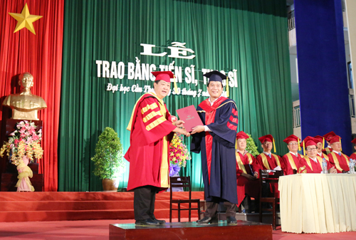
Tên đề tài: “Nghiên cứu hiệu quả điều hòa enzyme chuyển hóa glucose của lá xoài non (Mangifera indica L.) và rễ me keo (Pithecellobium dulce (Roxb.) Benhth.) trên chuột bệnh đái tháo đường”.
Tác giả: Nguyễn Thị Ái Lan, Khóa: 2015
Chuyên ngành: Công nghệ sinh học; Mã số: 62420201. Nhóm ngành: Khoa học sự sống.
Người hướng dẫn chính: PGS.TS. Đái Thị Xuân Trang - Trường Đại học Cần Thơ
- Tóm tắt nội dung luận án
Xoài (Mangifera indica L.) và me keo (Pithecellobium ducle (Roxb.) Benth) được trồng phổ biến ở Đồng bằng sông Cửu Long. Trong nghiên cứu này, khả năng chống oxy hóa, điều hòa hoạt động của các enzyme glucose-6-phosphatase, glucose-6-phosphate dehydrogenase và lactate dehydrogenase, bảo vệ tế bào min6 tụy tạng của cao chiết methanol lá xoài non và rễ me keo được nghiên cứu in vitro. Độc tính cấp của cao chiết lá xoài non và rễ me keo cũng được khảo sát trên chuột nhắt trắng. Khả năng chống tăng glucose huyết, rối loạn lipid huyết, chống xơ vữa động mạch, điều hòa enzyme glucose-6-phosphatase, glucose-6-phosphate dehydrogenase và lactate dehydrogenase in vivo của cao chiết lá xoài non và rễ me keo cũng được thực hiện trên mô hình chuột tăng glucose huyết. Bên cạnh đó, nghiên cứu còn đánh giá ảnh hưởng của cao chiết lá xoài non và rễ me keo đến cholesterol toàn phần, triglyceride, HDL-Cho, LDL-Cho và các chỉ số tim mạch (AI, CRI, CVRI). Mô hình chuột tăng glucose huyết cảm ứng bởi alloxan monohydrate được tiến hành khảo sát trong 28 ngày. Chuột tăng glucose huyết được chia thành 11 nhóm, mỗi nhóm gồm 6 con chuột có khối lượng và glucose huyết khác biệt không có ý nghĩa thống kê. Nghiệm thức I là nhóm đối chứng sinh lý (chuột bình thường không tiêm alloxan monohydrate hay uống bất kỳ loại thuốc nào). Nghiệm thức II, III là nhóm chuột bình thường uống 450 mg/kg khối lượng cao chiết lá xoài non hoặc rễ me keo. Nghiệm thức IV là nhóm đối chứng bệnh lý (chuột tăng glucose huyết không được điều trị). Nghiệm thức V là nhóm chuột tăng glucose huyết dùng thuốc biệt dược glucophage (108 mg/kg khối lượng). Các nghiệm thức tiếp sau là chuột tăng glucose huyết được điều trị bằng cao chiết lá xoài non hoặc rễ me keo (liều 150, 300, 450 mg/kg thể trọng/ lần x 2 lần/ ngày). Cao chiết lá xoài non hoặc rễ me keo được khảo sát khả năng gây độc tính cấp trên chuột nhắt trắng (Mus musculus L.) ở các nồng độ 1000, 2500 và 5000 mg/kg khối lượng in vivo.
Cao chiết lá xoài non và rễ me keo có chứa các hợp chất flavonoid, alkaloid, tannin, triterpenoid, glycoside. Cao chiết lá xoài non còn chứa thêm các chất courmarin và quinon; rễ me keo chứa thêm các chất phenol và saponin. Hàm lượng polyphenol tổng (total polyphenol content, TFC) của lá xoài non (335,06 ± 1,84 mg GAE/g cao chiết) cao hơn rễ me keo (246,5 ± 65,5 mgGAE/g cao chiết). Hàm lượng flavonoid toàn phần (total flavonoide content, TFC) của lá xoài non (432,86 ± 10,01 mg QE/g cao chiết) cũng cao hơn rễ me keo (427,4 ± 4,9 mg QE/g cao chiết). Hoạt tính chống oxy hóa được xác định bằng các phương pháp khác nhau như DPPH, ABTS+ và RP. Kết quả cho thấy lá xoài non và rễ me keo đều có hiệu quả chống oxy hóa nhưng đều thấp hơn chất chống oxy hóa chuẩn. Hiệu quả loại bỏ gốc tự do DPPH của lá xoài non (EC50= 27,6 ± 0,88 µg/mL) cao hơn rễ me keo (28,9 ± 0,80 µg/mL) và thấp hơn chất chuẩn trolox (4,26 µg/mL). Ngược lại, rễ me keo có khả năng khử sắt (69,3 ± 1,34 µg/mL) và trung hòa gốc tự do ABTS+ (23,8 ± 2,4 µg/mL) cao hơn lá xoài non ở cả hai phương pháp lần lượt là 321,4 ± 6,63 µg/mL và 45,7 ± 0,5 µg/mL; nhưng thấp hơn chất chuẩn BHA (30,1 µg/mL) trong phương pháp khử sắt và trolox (0,037 µg/mL) trong phương pháp ABTS+. Kết quả thực nghiệm khảo sát hiệu quả điều hòa hoạt động các enzyme chuyển hóa glucose cho thấy, LXN ức chế hoạt động G6Pase (IC50 = 80,4 µg/mL), G6PDH (IC50= 82,6 µg/mL) tốt hơn so với RMK (IC50 = 26,8 µg/mL, IC50 = 92,7 µg/mL). LXN (IC50 = 117,9 µg/mL) ức chế hoạt động enzyme LDH tốt hơn so với RMK (IC50 = 122,4 µg/mL). Nồng độ và thời gian tối ưu của tunicamycin để gây chết tế bào min6 tụy tạng là 5 µg/mL ở thời điểm 24 và 48 giờ. Cao chiết lá xoài non hoặc rễ me keo không gây độc tế bào đối với tế bào min6 trong 48 giờ ở tất cả nồng độ khảo sát từ 50-500 µg/mL. Nồng độ cao chiết lá xoài non hoặc rễ me keo có khả năng bảo vệ tế bào min6 khỏi sự chết do tác động của tunicamycin là 50 và 100 µg/mL khi khảo sát lần lượt ở thời gian 24 và 48 giờ.
Cao chiết lá xoài non (450 mg/kg) hoặc rễ me keo (150 mg/kg) được kết luận là có khả năng hạ glucose huyết, điều hòa lipid huyết và chống huyết khối ở chuột tăng glucose huyết. Kết quả quan sát cấu trúc mô bệnh học gan và thận chuột cho thấy, lá xoài non (450 mg/kg) hoặc rễ me keo (150 mg/kg) có khả năng cải thiện mô gan và thận của chuột tăng glucose huyết trở về tương đương với gan và thận ở chuột bình thường. Sau 14 ngày theo dõi, cao chiết lá xoài non và rễ me keo được chứng minh không gây ảnh hưởng bất thường nào đến các hành vi như lãnh cảm, tăng động, hệ hô hấp, nhịp tim và thông số AST, ALT của chuột trong suốt thời gian thí nghiệm. Bên cạnh đó, kết quả cho thấy lá xoài non hoặc rễ me keo ở các liều khảo sát không ảnh hưởng đến hành vi, sự tăng trọng bình thường của chuột. Các chỉ số về glucose huyết, huyết học, chức năng gan và thận ở nhóm chuột uống cao chiết lá xoài non hoặc rễ me keo ở các liều khảo sát đều tương đương với chuột bình thường. Điều này cho thấy lá xoài non hoặc rễ me keo không gây độc tính cấp trên chuột ở nồng độ khảo sát 5000 mg/kg. Cấu trúc mô học của gan và thận ở nhóm chuột uống cao chiết lá xoài non hoặc rễ me keo liều cao có ảnh hưởng đến cấu trúc mô gan và thận chuột bình thường. Tóm lại, cao chiết lá xoài non (450 mg/kg) và rễ me keo (150 mg/kg) có khả năng chống tăng glucose huyết, rối loạn chuyển hóa lipid, chống xơ vữa động mạch tốt nhất. Cao chiết lá xoài non hoặc rễ me keo có thể được sử dụng như một loại dược liệu hữu dụng trong điều trị tăng glucose huyết và ngăn ngừa biến chứng do sự tăng glucose huyết kéo dài gây ra.
- Những kết quả mới của luận án
Về hóa thực vật: đề tài đã khảo sát thành phần hóa thực vật và sơ bộ định lượng hàm lượng flavonoid và polyphenol tổng. Các dữ liệu này có thể đóng góp thêm các thông tin cho việc sử dụng lá xoài non và rễ me keo trong y học cổ truyền.
Về mặt cơ chế sinh học: để tài đã khảo sát tác động cùa các cao chiết lá xoài non và rễ me keo đối với hoạt động của 03 enzyme G6Pase, G6PDH, LDH. Đây là những enzyme liên quan trực tiếp đến cơ chế chuyển hóa và hấp thụ glucose. Đồng thời đã khảo sát tác động bảo vệ của cáo chiết là xoài non và rễ me keo đối với tế bào tụy tạng min6.
Đề tài đã đánh giá tác dụng chống tăng glucose huyết và rối loạn lipid huyết của cao chiết lá xoài non và rễ me keo trên mô hình chuột nhắt trắng gây bệnh đái tháo đường bằng alloxan monohydrate. Các tác động trên mô học gan, thận và hiệu quả điều hòa hoạt động của G6Pase, G6PDH và LDH in vivo cũng được đề tài đánh giá.
- Các ứng dụng trong thực tiễn
Lần đầu tiên các kết quả về tác dụng của cao chiết lá xoài non và rễ me keo đối với các enzyme G6Pase, G6PDH và LDH được nghiên cứu và làm rõ ở cấp độ in vitro và in vivo. Tác động dược lý bảo vệ tế bào tụy tạng min6 và các tác dụng trên gan, thận được trình bày, giúp cung cấp và đóng góp dữ liệu khoa học về loài dược liệu này. Đồng thời gợi ý tiềm năng của dược liệu trong nghiên cứu có thể được ứng dụng hỗ trợ và điều trị bệnh đái tháo đường trong tương lai.
- Summary of doctoral thesis
Mangifera indica L. and Pithecellobium dulce (Roxb.) Benth. are popular in Mekong delta. So in this research, the antioxidant role, regulating glucose-6-phosphatase (G6Pase), glucose-6-phosphate dehydrogenase (G6PDH) and lactate dehydrogenase (LDH), protective effect min6 pancreatic β-cells, antidyslipidemic of Mangifera indica L. young leaves and Pithecellobium ducle (Roxb.) Benth. root were investigated in vitro. Acute toxicity studies with MIL or PDR were performed in experimental mice. The present study was also conducted to elucidate the anti-hyperglycemic, anti- dyslipidemia, anti- atherosclerosis. The activities of key carbohydrate metabolism enzyme comprising G6Pase, G6PDH and LDH in liver and muscle of diabetic mice were determined. The in vivo experiment, mice were orally treated with three different single doses 1000, 2500, 5000 mg/kg of the MIL or DPR extract and screened for signs of acute toxicity. This study was assessed by determining its effect on blood glucose, body weight, total cholesterol, triglyceride, HDL- Cho, LDL- Cho, atherogenic index (AI, CRI, CVRI) incompared with those of controls. Alloxan monohydrate (AM) - induced diabetic male Mus musculus L. mice were used as experimental models for a period of 28 days. The experiment was designed to determine effect of MIL or PDR extract on diabetic mice. The mice were allowed to acclimated to the laboratory environment for 48 hours, distributed into eleven groups according to the similar weights and glucose blood with six animals in each group (n=6). Group I normal control consisted of normal mice that neither injected AM nor any drug. Group II and III were taken MIL or PDR extract (450 mg/kg body weight). Group IV served as negative control (diabetic control). Group V was given standard drug, glucophage (170 mg/ kg body weight as possitive control. Next groups, diabetic mice were treated with extract of MIL or PDR (150, 300, 450 mg/kg body weight/ twice a day).
Hyperglycermia was resulted in the generation of free radicals. So the in vitro studies including phytochemical screening of methanol extracts of MIL and PDR were examined the presence of bioactive compounds. The antioxidant activities of MIL and PDR were demonstrated by employing established in vitro systems, which included radical scavenging DPPH, ABTS·+ and ferric reducing. Pancreatic beta cell line closely associated with to diabetes mellitus. So they become weak or die, insulin secretion will be affected immediately. Protective effect of MIL or PDR extract against endoplasmic reticulum (ER) stress was conducted in min6 pancreatic β-cells in vitro. The min6 cell line was cultured with 5 µg/mL tunicamycin added in 24 hours of exposure and 5% CO2 to induce ER stress and cell death. Cytotoxicity effect of MIL or PDR extract on min6 cells was observed concentration from 50 to 500 µg/mL. To investigate the effect of MIL or PDR extract on ER stress, min6 cell were treatment tunicamycin with extract (50 - 500 µg/mL) for 48 hours at 37oC in a 5% CO2 humidified incubator. The first result show that phytochemical investigation on the MIL and PDR methanol extracts revealed the presence of various phytoconstituents such as flavonoids, alkaloids, tannins, triterpenoids and glycosides. MIL also contains phenols, coumarins and quinons while PDR contains saponins. The total polyphenol of MIL extract (335.06 ± 0.05 mg GAE/g extract) is higher than amount of total polyphenols of PDR (246.5 ± 65.5 mg GAE/g extract). Total flavonoid content of MIL (432.86 ± 10.01 mg QE/g extract) is also higher than TFC of PDR (427.4 ± 4.9 mg QE/g extract). In addition, the antioxidant effects of the MIL and PDR indicate that both MIL and PDR extract against oxidation effect but there are lower than standard. In DPPH assay, the anti-oxidation of MIL (EC50= 27.6 µg/mL) is higher than PDR (EC50= 67.9 µg/mL) and lower than Trolox standard (EC50= 4.26 µg/mL). In Fe2+ chelating and ABTS assay, the anti-oxidation of PDR extract (67.9 µg/mL, 7.03 µg/mL), is higher than MIL extract (45.7±0.50 µg/mL; 12.11±1.15 µg/mL, respectively) but lower than BHA standard (30.1 µg/mL) and Trolox standard (0.037 µg/mL). Addition, the second result indicate that at concentration 5 µg/mL, tunicamycin had significant induced apoptosis after 24 hours. MIL and PDR did not cause min6 cell to die for 48 hours. The extract of MIL (50 µg/mL) or PDR (100 µg/mL) can protect min6 cells against cell death with ER stress respectively in 24 and 48 hours. Next, the result screening of controlling glucose enzyme metabolism activities of extracts depicted the effect of MIL on G6Pase (IC50 = 42.9 µg/mL), G6PDH (IC50 = 82.6 µg/mL) activities is better than PDR (IC50 = 56 µg/mL, 92.7 µg/mL). In contrast, the effect of PDR extract on LDH(IC50 = 122.6 µg/mL) activity is higher than MIL (IC50 = 142.6 µg/mL, respectively).
The results showed that the methanol extract of MIL or PDR showed significantly antidiabetic and antihyperlipidemic activities as evidenced by a significant decrease in plasma glucose, TC, TG, LDL- Cho levels with elevation of HDL- Cho level and diminution of the atherogenic index in diabetic mice. Observation of the microscopic cross-section of liver tissues revealed that mice treated with MIL or PDR extract had significantly improvement in liver tissues compared to those of the non-treated control group. Further more, throughout the 14 days observation acute period, no abnormal changes attributable to the treatment tunicamycin were noticed in behavior such as apathy, hyperactivities, morbidity, respirations and hear rate etc. Besides, the body weight, blood glucose, AST and ALT were not significant changed after the period of experiment. Observation of the microscopic cross section of liver and tissues revealed that mice treated with high extracts had not change when compared in those of the control group. In conclution, the MIL or PDR hold anti diabetic, antihyperlipidemic activity and antiatherogenicity in diabetic mice. They can be utilized as useful remedy for alleviation of diabetes and its complication.
- Innoative contribute of the thesis
Regarding phytochemicals: the thesis investigated the phytochemical composition and preliminary quantitative determination of total flavonoids and polyphenols. These data may contribute additional information for the use of young Mangifera indica L. leaves and Pithecellobium ducle (Roxb.) Benth. root in traditional medicine.
Regarding biological mechanism: studying the effects of extracts of young Mangifera indica L. leave and Pithecellobium ducle (Roxb.) Benth. on the activities of 03 G6Pase, G6PDH, LDH enzymes. These enzymes directly involved in glucose metabolism and absorption. The protective effects of young Mangifera indica L. leave and Pithecellobium ducle (Roxb.) Benth. extract on pancreatic cells of min6 were investigated.
The study evaluated the antihyperglycemic and dyslipidemic effects of young Mangifera indica L. leave and Pithecellobium ducle (Roxb.) Benth. extract on a model of mice causing diabetes with alloxan monohydrate. The effects on hepatic and renal histology and the regulatory effects of G6Pase, G6PDH and LDH in vivo were also evaluated.
- Practical significance
The thesis provides new scientific information about the effects of young mango leaf extract and acacia root extract on enzymes G6Pase, G6PDH and LDH in vitro and in vivo. The protective pharmacological effects of min6 pancreatic cells and the effects on the liver and kidneys are presented, helping to provide and contribute to scientific data on this medicinal species. besides that suggesting the potential of medicinal herbs in research could be applied to support and treat diabetes in the future.
- Xem chi tiết nội dung luận án
- Xem thông tin đăng tải tại Website Bộ giáo dục và Đào tạo. (Nhập tên NCS vào ô tìm kiếm)
Tin mới nhất
Hướng dẫn HVCH nhập Kế hoạch học tập lên Hệ thống quản lý


Số lượt truy cập